4.Identify the anatomy and electrical conduction pathway of the heart.
5.Describe the proper procedure for obtaining a 12-lead EKG, including patient preparation, lead placement, and artifact prevention.
6.Interpret basic EKG waveforms, intervals, and segments, and recognize common arrhythmias.
7.Explain the medical assistant's role in cardiovascular testing, including Holter monitor application, stress testing, and patient education.
8.Apply safety, infection control, and legal standards (OSHA Bloodborne Pathogens Standard, HIPAA Privacy Rule, CDC Standard Precautions) during all cardiovascular procedures.
1.1 Anatomy and Physiology of the Heart
The heart is a four-chambered muscular pump located in the mediastinum. The upper chambers are the atria (right and left), and the lower chambers are the ventricles (right and left). The right side of the heart pumps deoxygenated blood to the lungs (pulmonary circulation), while the left side pumps oxygenated blood to the body (systemic circulation).
The coronary arteries (left main, left anterior descending, left circumflex, right coronary artery) supply the heart muscle itself with oxygenated blood. Blockage of these vessels leads to myocardial infarction (heart attack).
Electrical Conduction System
The heart has its own electrical system that triggers mechanical contraction. The sequence is:
16.Sinoatrial (SA) node – the primary pacemaker, located in the right atrium. Fires at 60–100 beats per minute (bpm).
17.Atrioventricular (AV) node – located at the junction of the atria and ventricles. Delays the impulse to allow atrial contraction to finish filling the ventricles.
18.Bundle of His – conducts the impulse from the AV node to the interventricular septum.
19.Right and Left Bundle Branches – carry the impulse down the septum to the ventricular walls.
20.Purkinje fibers – spread the impulse upward through the ventricular myocardium, causing ventricular contraction.
If the SA node fails, the AV node takes over at 40–60 bpm. If both fail, the Purkinje fibers fire at 20–40 bpm (a life-threatening emergency).
1.2 The 12-Lead EKG: Principles and Preparation
An electrocardiogram (EKG or ECG) records the heart's electrical activity through electrodes placed on the skin. The 12-lead EKG provides 12 different views (leads) of the heart's electrical activity from 10 electrodes. It is used to detect arrhythmias, ischemia, infarction, electrolyte imbalances, and chamber enlargement.
Patient Preparation
Verify patient identity using two identifiers (name and date of birth) per facility policy.
Explain the procedure: it is painless, takes 5–10 minutes, and requires lying still.
Ask the patient to remove all jewelry, watches, and metal objects from the chest and wrists.
The chest must be bare. For female patients, provide a gown that opens in the front. Do not lift or remove the gown without permission.
Shave excessive chest hair if it prevents electrode adhesion (use a disposable razor).
Clean the skin with alcohol or an abrasive pad to remove oils and dead skin cells. Allow the skin to dry completely before applying electrodes.
Position the patient supine (lying flat on the back). If the patient cannot tolerate this, use semi-Fowler's position and document it, as position changes can alter the EKG.
Electrode Placement (Standard 12-Lead)
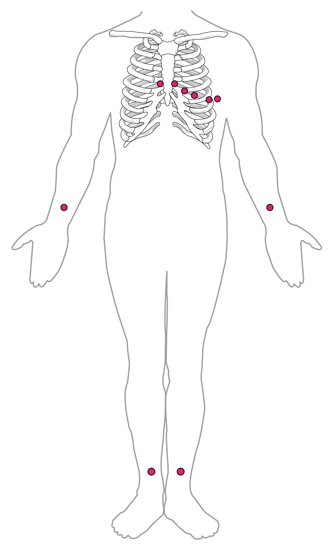
There are 4 limb electrodes and 6 chest (precordial) electrodes.
Limb leads (placed on the fleshy parts of the arms and legs, not on bone):
RA (Right Arm) – white
LA (Left Arm) – black
RL (Right Leg) – green (ground/reference)
LL (Left Leg) – red
Chest leads (V1–V6):
V1 – 4th intercostal space, right sternal border (red)
V2 – 4th intercostal space, left sternal border (yellow)
V3 – midway between V2 and V4 (green)
V4 – 5th intercostal space, midclavicular line (blue)
V5 – 5th intercostal space, anterior axillary line (orange)
V6 – 5th intercostal space, midaxillary line (purple)
Memory aid for chest leads: "White on right, smoke over fire" (for limb leads: white on right arm, black on left arm, green on right leg, red on left leg). For chest leads: "V1 to V6, right to left, bottom to top."
Artifact Prevention
Artifact is unwanted interference on the EKG tracing. Common causes and fixes:
Muscle tremor (somatic tremor) – patient shivering or tense. Fix: reassure patient, keep them warm, ask them to relax arms and legs.
AC interference (60-cycle interference) – electrical devices nearby (IV pumps, monitors). Fix: unplug nearby devices, ensure electrodes are not crossed.
Broken or crossed leads – check color coding and connections.
1.3 EKG Waveforms, Intervals, and Segments
The EKG tracing consists of waves, segments, and intervals that correspond to electrical events.
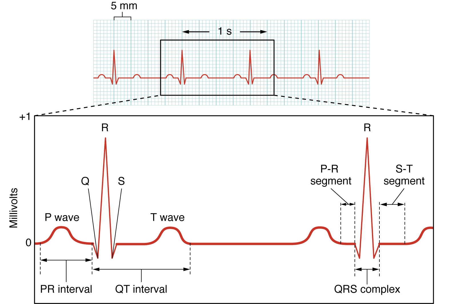
P wave – atrial depolarization (contraction). Normally upright, small, rounded, and < 0.12 seconds wide.
PR interval – from the start of the P wave to the start of the QRS complex. Normal: 0.12–0.20 seconds (3–5 small squares). Represents the delay at the AV node.
QRS complex – ventricular depolarization. Normal duration: 0.06–0.10 seconds (1.5–2.5 small squares). The Q wave is the first negative deflection, R is the first positive deflection, S is the negative deflection after R.
ST segment – from the end of the QRS to the start of the T wave. Represents early ventricular repolarization. Should be flat (isoelectric). Elevation or depression indicates ischemia or injury.
T wave – ventricular repolarization. Normally upright and rounded.
QT interval – from the start of the QRS to the end of the T wave. Represents total ventricular electrical activity. Prolonged QT can lead to arrhythmias.
U wave – a small wave after the T wave, often seen in hypokalemia.
EKG Paper and Rate Calculation
EKG paper runs at 25 mm/second. Each small square = 1 mm = 0.04 seconds. Each large square (5 small squares) = 0.20 seconds. Five large squares = 1 second.
To calculate heart rate:
Method 1 (6-second strip): Count the number of QRS complexes in a 6-second strip (30 large squares) and multiply by 10.
Method 2 (large square method): Divide 300 by the number of large squares between two consecutive R waves (R-R interval). For example, if there are 4 large squares between R waves, rate = 300 ÷ 4 = 75 bpm.
Method 3 (small square method): Divide 1500 by the number of small squares between R waves.
1.4 Common Arrhythmias and EKG Findings
Normal Sinus Rhythm (NSR): Rate 60–100 bpm, regular rhythm, one P wave before each QRS, normal PR and QRS intervals.
Sinus Bradycardia: Rate < 60 bpm, regular rhythm, normal P waves. Causes: athletes, sleep, hypothyroidism, medications. Symptomatic bradycardia may require treatment.
Sinus Tachycardia: Rate > 100 bpm, regular rhythm, normal P waves. Causes: exercise, fever, anxiety, dehydration, hyperthyroidism.
Atrial Fibrillation (A-fib): Irregularly irregular rhythm, no distinct P waves (replaced by fibrillatory "f" waves), normal QRS. High risk of stroke; patients often take anticoagulants.
Atrial Flutter: "Sawtooth" flutter waves (F waves) at 250–350 bpm, with a regular or irregular ventricular response.
Premature Ventricular Contraction (PVC): A wide, bizarre QRS complex occurring early, followed by a compensatory pause. Occasional PVCs are benign; frequent or multifocal PVCs may indicate heart disease.
Ventricular Tachycardia (V-tach): Wide QRS complexes at a rate > 100 bpm, regular. A life-threatening emergency; may progress to V-fib.
Ventricular Fibrillation (V-fib): Chaotic, irregular, no identifiable P waves or QRS complexes. No cardiac output – cardiac arrest. Requires immediate CPR and defibrillation per AHA CPR guidelines.
Asystole: Flatline (no electrical activity). Confirm in two leads. Requires CPR and epinephrine; do not defibrillate.
ST segment changes:
ST elevation – indicates acute myocardial injury (STEMI). Emergency.
ST depression – indicates myocardial ischemia (often seen during stress tests).
1.5 Holter Monitor and Event Monitor
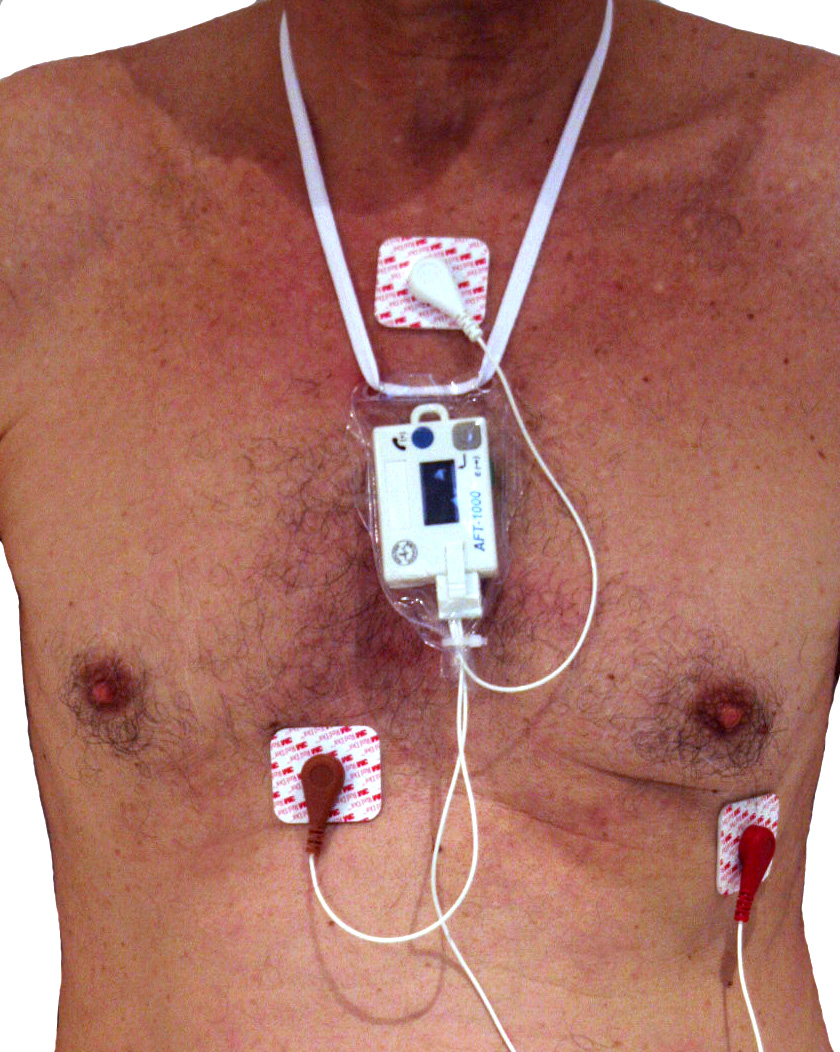
A Holter monitor is a portable device that continuously records the heart's rhythm for 24–48 hours. It is used to detect intermittent arrhythmias, palpitations, or syncope that a standard EKG may miss.
Medical Assistant's Role
Explain the purpose and duration of the test.
Prepare the skin (clean, dry, shave if needed) and apply electrodes (typically 3–5 leads) to the chest.
Connect the electrodes to the monitor, insert a fresh battery, and verify the device is recording.
Instruct the patient to:
Keep a diary of symptoms (time, activity, symptoms felt).
Press the event button on the monitor when symptoms occur.
Avoid showering, bathing, or swimming (monitor is not waterproof).
Avoid magnets, metal detectors, and high-voltage areas.
Continue normal daily activities to provoke symptoms.
Document the application time, electrode sites, and patient education in the chart.
An event monitor is similar but is worn for weeks and records only when the patient activates it or when an arrhythmia is automatically detected.
1.6 Stress Testing (Exercise Tolerance Test)
A stress test evaluates the heart's response to exercise. It is used to diagnose coronary artery disease, assess exercise tolerance, and evaluate treatment effectiveness. The patient walks on a treadmill or pedals a stationary bike while the EKG, blood pressure, and heart rate are monitored.
Medical Assistant's Role
Verify the patient has followed pre-test instructions (no caffeine, no smoking, no food for 2–4 hours, and physician instructions regarding medications).
Obtain baseline vital signs and a resting 12-lead EKG.
Apply electrodes and attach the EKG monitor and blood pressure cuff.
Monitor the patient for symptoms: chest pain, dyspnea, dizziness, excessive fatigue.
Monitor vital signs and EKG during and after the test.
Stop the test if the patient experiences severe symptoms, significant ST changes, dangerous arrhythmias, or a drop in blood pressure.
Assist the patient to a reclining position during recovery and monitor until vital signs return to baseline.
Contraindications to stress testing: acute myocardial infarction, unstable angina, uncontrolled arrhythmias, severe aortic stenosis, acute heart failure.
1.7 Infection Control and Safety in Cardiovascular Testing
Follow CDC Standard Precautions for all patients: perform hand hygiene before and after patient contact, wear gloves when there is potential contact with blood or body fluids, and dispose of sharps in puncture-resistant containers.
The OSHA Bloodborne Pathogens Standard requires the use of personal protective equipment (PPE) and proper disposal of contaminated waste. If skin is exposed to blood, wash immediately with soap and water.
Clean and disinfect EKG cables, electrodes, and equipment between patients. Use single-use electrodes and disposable razors.
Ensure the EKG machine is properly grounded to prevent electrical shock.
Maintain patient privacy per the HIPAA Privacy Rule: do not discuss results in hallways, close curtains, and only share information with authorized personnel.
1.8 Patient Education and Documentation
Explain each procedure in simple terms, addressing anxiety and answering questions.
For EKG: "This test records your heart's electrical activity. It does not send electricity through you."
For Holter monitor: "Wear the monitor for 24 hours. Keep your diary and press the button if you feel symptoms."
Document: date and time of procedure, patient preparation, lead placement, any artifacts or difficulties, patient tolerance, and the tracing (or a copy) in the chart. Note any symptoms reported by the patient.
Report abnormal findings (e.g., ST elevation, dangerous arrhythmias) to the provider immediately. Do not interpret or diagnose – the medical assistant's role is to obtain a quality tracing and alert the provider.
Common Exam Traps
127.Confusing lead placement colors. Students often mix up the limb leads. Remember: White on Right, Black on Left, Green on Right Leg (like a traffic light: red, yellow, green – but red is on the left leg). Chest leads V1–V6 go from right sternum to left midaxillary line, not left to right.
128.Calculating heart rate incorrectly. The 300 method uses large squares between R waves. If you count small squares, you must divide 1500 by that number. Do not divide 300 by small squares – that is a common error.
129.Thinking the SA node fires at 40–60 bpm. That is the AV node rate. The SA node fires at 60–100 bpm. The Purkinje fibers fire at 20–40 bpm.
130.Confusing atrial fibrillation with ventricular tachycardia. A-fib has an irregularly irregular rhythm with no P waves but narrow QRS complexes. V-tach has wide QRS complexes and is regular. If you see a wide-complex tachycardia, think V-tach, not A-fib.
131.Forgetting the PR interval normal range. It is 0.12–0.20 seconds (3–5 small squares). A PR interval longer than 0.20 seconds indicates first-degree AV block. A short PR interval (< 0.12) may indicate Wolff-Parkinson-White syndrome.
132.Applying electrodes over bone or hair. Electrodes must be on fleshy areas. Over bone (clavicle, sternum, ribs) or hair causes artifact and poor tracing.
133.Telling the patient to shower with a Holter monitor. Holter monitors are not waterproof. Patients must avoid showering, bathing, and swimming. Only a sponge bath is allowed.
134.Stopping a stress test for mild fatigue. The test is stopped for severe symptoms, dangerous arrhythmias, ST elevation, or a drop in blood pressure – not for mild, expected fatigue.
135.Confusing ST elevation with ST depression. ST elevation = acute injury (STEMI, emergency). ST depression = ischemia (often stable, but still report). Both are abnormal.
136.Forgetting to document. Even if you performed the EKG perfectly, if you did not document the time, patient tolerance, and any issues, the chart is incomplete. Documentation is a legal and professional requirement.
Summary
The medical assistant plays a critical role in cardiovascular testing: preparing the patient, obtaining a high-quality 12-lead EKG, applying Holter monitors, assisting with stress tests, and recognizing when to alert the provider. Mastery of lead placement, rate calculation, waveform interpretation, and safety protocols is essential for the NHA CCMA exam and for safe clinical practice. Always follow CDC Standard Precautions, OSHA Bloodborne Pathogens Standard, and HIPAA Privacy Rule. Remember: your job is to obtain and document accurate data – not to diagnose. When in doubt, report to the provider.
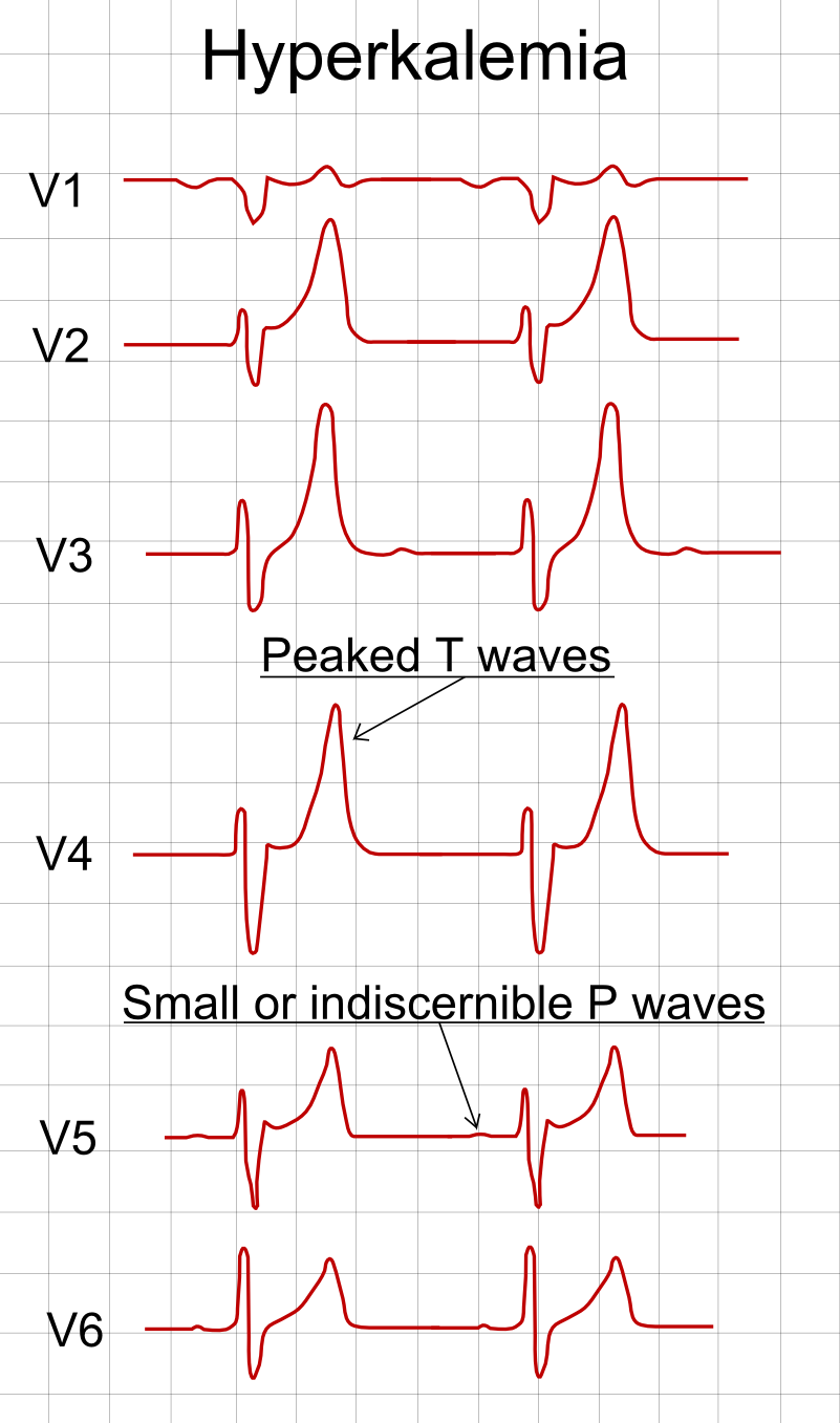
Precordial lead placement
Precordial lead placement
Holter monitor
Holter monitor
Standard 12-lead ECG electrode placement
Standard 12-lead ECG electrode placement
ECG waveform: P-QRS-T waves, intervals and segments
ECG waveform: P-QRS-T waves, intervals and segments