Differentiate between CLIA-waived, moderate-complexity, and high-complexity tests
Identify the correct order of draw for venipuncture and capillary collection
List the components of a standard urinalysis and their clinical significance
Apply OSHA Bloodborne Pathogens Standard precautions during specimen handling
Recognize the proper procedure for glucose testing, hemoglobin testing, and rapid strep testing
Calculate and interpret basic hematocrit and hemoglobin values
Identify pre-analytical, analytical, and post-analytical errors in testing
Select the correct anticoagulant for common laboratory tests
Explain the chain of custody procedure for forensic specimens
Apply HIPAA Privacy Rule requirements to laboratory results and patient communication
1.1 Overview of Point of Care Testing (POCT)
Point of care testing refers to diagnostic testing performed at or near the site of patient care, providing rapid results that guide immediate clinical decisions. The Clinical Laboratory Improvement Amendments (CLIA) regulate all laboratory testing performed on humans in the United States, regardless of location. CLIA categorizes tests by complexity: waived, moderate, and high.
Waived tests are simple procedures with a low risk of error. They require minimal training and are cleared by the FDA for home use or are so simple that the likelihood of erroneous results is negligible. Common waived tests include:
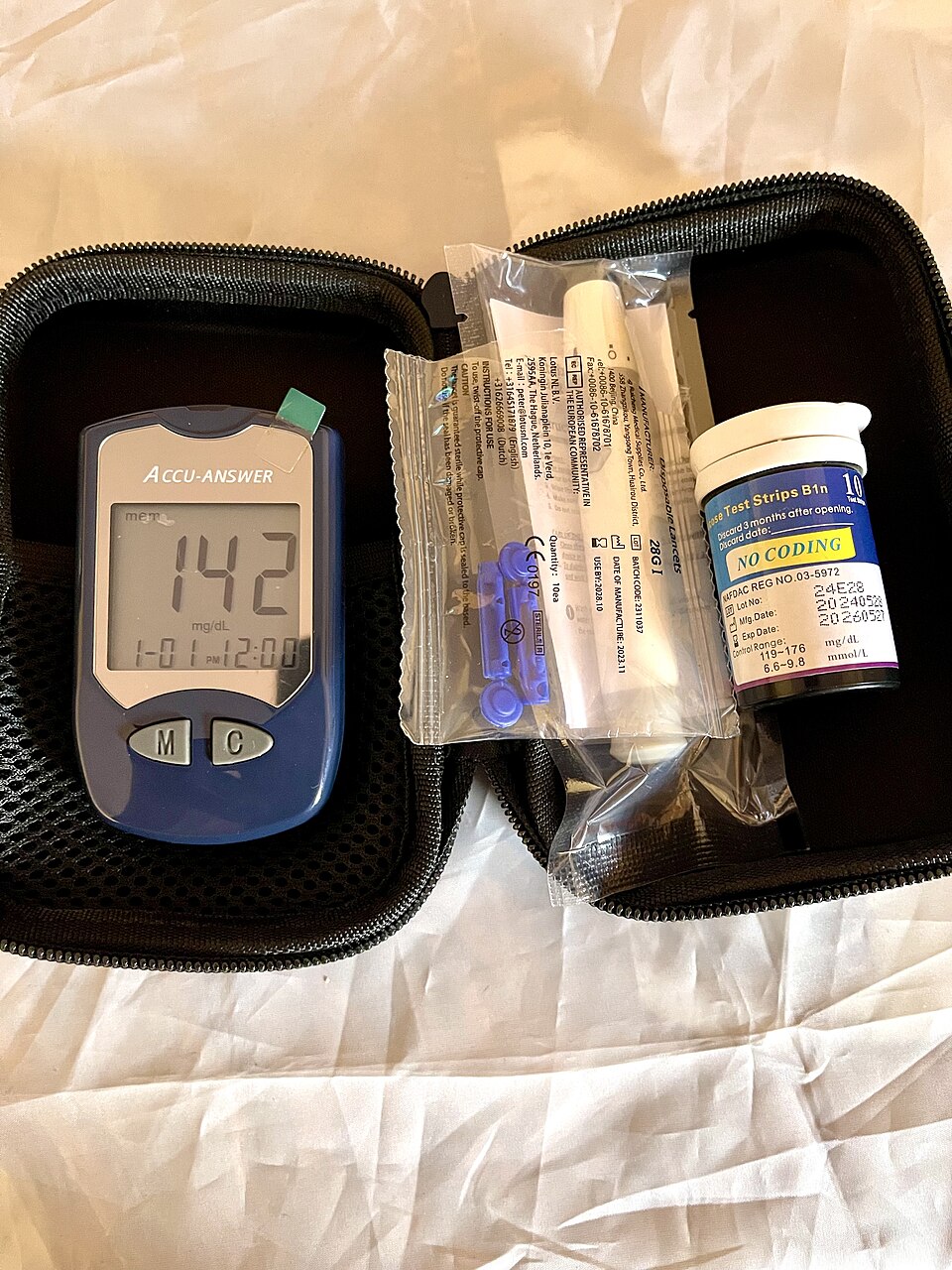
Blood glucose monitoring
Urine pregnancy tests
Rapid strep A tests
Influenza A/B tests
Fecal occult blood tests
Hemoglobin (Hgb) via portable analyzers
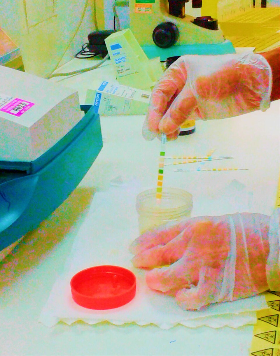
Urine dipstick analysis
Moderate-complexity tests require more training, quality control, and documentation. Examples include:
Complete blood count (CBC) using automated analyzers
Coagulation panels (PT/INR)
Basic metabolic panels
High-complexity tests include cytology, histopathology, and advanced microbiology. These are performed in reference laboratories by specialized personnel.
The medical assistant (MA) is typically responsible for waived testing. However, the MA must still follow manufacturer instructions exactly, perform quality control checks, document results, and maintain the testing log. CLIA requires that the facility hold a valid Certificate of Waiver, and the MA must never modify the test procedure, use expired reagents, or skip calibration steps.
1.2 Safety and Standard Precautions in the Laboratory
All laboratory work falls under the OSHA Bloodborne Pathogens Standard. This standard requires employers to provide personal protective equipment (PPE), hepatitis B vaccination, and annual training. The MA must treat all blood and body fluids as potentially infectious, regardless of the patient's known status.
CDC Standard Precautions apply to all patient contact and specimen handling. Key components include:
Hand hygiene before and after every patient interaction
Gloves for any procedure involving blood, body fluids, or mucous membranes
Goggles or face shields when splashing is possible
Gowns or aprons for procedures with high splash risk
Safe sharps disposal in puncture-resistant, leak-proof containers
Never recapping needles, bending, or breaking them
Needlestick protocol: If a needlestick occurs, the MA must immediately wash the area with soap and water, report the incident to the supervisor, and seek medical evaluation. The exposure must be documented per facility policy. Do not squeeze the wound, and do not apply bleach or harsh chemicals.
Specimen transport: All specimens must be placed in leak-proof bags with a biohazard label. The requisition slip must never be placed inside the specimen bag—it goes in the outer pocket to prevent contamination.
Spill cleanup: For a blood spill, the MA should wear gloves, cover the spill with absorbent material, apply an EPA-registered disinfectant (e.g., 10% bleach solution), allow appropriate contact time, then wipe up and dispose of materials in biohazard waste.
1.3 Capillary Puncture (Microcollection)
Capillary puncture is used for infants, toddlers, elderly patients with fragile veins, patients with severe burns, and for tests requiring only small volumes (e.g., glucose, hemoglobin, newborn screening).
Sites:
Adults and children over 1 year: lateral aspect of the distal phalanx of the middle or ring finger. Avoid the index finger (more calloused) and the thumb (more arterial supply). The puncture should be perpendicular to the fingerprint ridges.
Infants under 1 year: medial or lateral plantar surface of the heel (the outer edges). Never use the center of the heel (calcaneus bone risk) or the arch.
Procedure:
51.Warm the site with a warm compress for 3–5 minutes to increase blood flow.
52.Clean the site with an alcohol wipe and allow it to dry completely (alcohol causes hemolysis and stinging).
53.Use a sterile lancet (auto-retracting preferred). Puncture depth should be ≤ 2.0 mm for infants, ≤ 2.4 mm for adults.
54.Wipe away the first drop of blood (contains tissue fluid).
55.Collect blood into microcollection tubes. Do not squeeze or "milk" the finger—this causes hemolysis and tissue fluid contamination. Gentle pressure is acceptable.
56.Cap the tube, invert it the required number of times, and label it.
Order of draw for capillary specimens (per CLSI standards):
58.Blood gas (heparinized capillary tube)
59.EDTA (lavender)
60.Other additive tubes
61.Serum tubes (red or SST)
Common errors: milking the site, using alcohol that is still wet, insufficient blood volume, and incorrect tube order.
1.4 Venipuncture and Order of Draw
Venipuncture is the collection of blood from a vein, typically the antecubital fossa (median cubital vein first choice, cephalic second, basilic third). The basilic vein is close to the brachial artery and median nerve—use with caution.
68.Verify the patient's identity using two identifiers (name and date of birth, or name and medical record number). Ask the patient to state their name—do not call it out.
69.Position the patient with the arm extended and supported.
70.Apply the tourniquet 3–4 inches above the puncture site. It should be tight enough to occlude venous flow but not arterial flow. Leave it on for no more than 1 minute.
71.Palpate for a vein. The vein should feel bouncy and refill after pressure.
72.Clean the site with 70% isopropyl alcohol in a circular motion, from center outward. Allow to dry.
73.Anchor the vein with the thumb 1–2 inches below the site.
74.Insert the needle at a 15–30 degree angle, bevel up. You will feel a "pop" when entering the vein.
75.Push the tube onto the holder. Blood should flow. If not, adjust the needle slightly.
76.Fill tubes in the correct order. Remove the tourniquet as soon as blood flows (or after the first tube).
77.Place gauze over the site and withdraw the needle. Apply pressure for 2–3 minutes (5 minutes for patients on anticoagulants).
78.Label tubes at the bedside, in front of the patient.
79.Dispose of the needle immediately into the sharps container.
Order of draw (critical to memorize):
81.Blood culture bottles (sterile, no additive)
82.Light blue (sodium citrate—for coagulation tests)
83.Red (no additive, serum) or SST/gold (serum separator gel)
Why order matters: If EDTA (lavender) contaminates the light blue tube, the citrate will bind calcium and produce false coagulation results. If heparin contaminates the red tube, it interferes with serum chemistry tests. The order ensures that additive carryover does not affect test accuracy.
Anticoagulant functions:
EDTA: chelates calcium, preventing clotting. Used for hematology.
Sodium citrate: binds calcium. Used for PT/INR, aPTT.
Heparin: activates antithrombin III. Used for chemistry and arterial blood gases.
Sodium fluoride: inhibits glycolysis (preserves glucose). Used for glucose and lactate.
Hematoma (needle through the vein, insufficient pressure after removal)
Clotted specimen (tubes not inverted enough)
Incorrect tube order (cross-contamination)
1.5 Hematology Tests
Hemoglobin (Hgb): Measures the oxygen-carrying protein in red blood cells. Normal ranges: males 13.5–17.5 g/dL, females 12.0–15.5 g/dL. Low hemoglobin indicates anemia; high may indicate polycythemia or dehydration.
Hematocrit (Hct): The percentage of blood volume occupied by red blood cells. Normal: males 38.8–50%, females 34.9–44.5%. The hematocrit is approximately 3× the hemoglobin value.
Microhematocrit procedure:
103.Fill a capillary tube with blood (from EDTA tube or capillary puncture).
104.Seal one end with clay.
105.Centrifuge for 5 minutes at 10,000–12,000 rpm.
106.Read using a microhematocrit reader. The buffy coat (white cell layer) is not included in the reading.
White blood cell (WBC) count: Normal 4,500–11,000/mm³. Elevated WBCs indicate infection, inflammation, or leukemia. Decreased WBCs may indicate viral infection, bone marrow suppression, or autoimmune disease.
Platelet count: Normal 150,000–450,000/mm³. Low platelets (thrombocytopenia) increase bleeding risk; high platelets (thrombocytosis) increase clotting risk.
Erythrocyte sedimentation rate (ESR): Measures inflammation. Blood is placed in a vertical tube, and the distance red cells fall in one hour is measured. Normal: males 0–15 mm/hr, females 0–20 mm/hr. Elevated ESR indicates inflammation, infection, or autoimmune disease.
1.6 Glucose Testing
Fasting blood glucose: Normal 70–99 mg/dL. Prediabetes: 100–125 mg/dL. Diabetes: ≥ 126 mg/dL on two occasions.
Random glucose: Normal < 140 mg/dL. ≥ 200 mg/dL with symptoms indicates diabetes.
Oral glucose tolerance test (OGTT): Patient fasts, then drinks 75 g of glucose. Blood is drawn at baseline, 1 hour, and 2 hours. A 2-hour value ≥ 200 mg/dL indicates diabetes. Used for gestational diabetes screening (50 g load, 1-hour draw).
Hemoglobin A1c: Reflects average glucose over 2–3 months. Normal < 5.7%. Prediabetes: 5.7–6.4%. Diabetes: ≥ 6.5%. This test does not require fasting.
POCT glucose procedure:
117.Verify the test strips are not expired and are coded to the meter.
118.Perform quality control (liquid control or electronic check) per manufacturer instructions.
119.Clean the patient's finger with alcohol and allow to dry.
120.Puncture the lateral fingertip, wipe the first drop, and apply the second drop to the strip.
121.Read the result and document it in the patient's record, including the time, value, and any relevant notes (e.g., fasting or postprandial).
122.Dispose of the lancet in the sharps container.
Critical error: Using expired strips, not coding the meter, or applying blood to a strip that has been exposed to humidity. Always store strips in their original container with the lid tightly closed.
1.7 Urinalysis
A complete urinalysis has three components: physical, chemical, and microscopic.
Physical examination:
Color: Normal is pale yellow to amber (urochrome). Red/brown suggests blood, beets, or porphyria. Dark yellow suggests dehydration. Cloudy suggests infection or phosphate crystals.
Clarity: Normal is clear. Turbidity indicates cells, bacteria, or crystals.
Specific gravity: Normal 1.005–1.030. Measures urine concentration. Fixed at 1.010 indicates renal failure.
Chemical examination (dipstick):
pH: Normal 4.5–8.0. Acidic urine (low pH) with uric acid stones; alkaline urine with struvite stones or urinary tract infection (UTI) from urea-splitting organisms.
Protein: Normal is negative or trace. Persistent protein indicates glomerular damage (nephrotic syndrome, diabetes).
Glucose: Normal is negative. Positive glucose indicates diabetes or renal glycosuria.
Ketones: Normal is negative. Positive ketones indicate fat metabolism (diabetes ketoacidosis, starvation, high-protein diet).
Blood: Normal is negative. Positive blood (hematuria) indicates infection, stones, trauma, or glomerulonephritis.
Leukocyte esterase: Normal is negative. Positive indicates pyuria (UTI).
Nitrite: Normal is negative. Positive indicates gram-negative bacteria (E. coli) that convert nitrate to nitrite.
Bilirubin: Normal is negative. Positive indicates liver disease or biliary obstruction.
Urobilinogen: Normal is 0.1–1.0 mg/dL. Elevated indicates hemolysis or liver disease.
Microscopic examination: Performed on centrifuged urine sediment. Findings include:
RBCs: > 3 per high-power field (HPF) is abnormal.
WBCs: > 5 per HPF indicates infection.
Casts: Hyaline casts are normal; red cell casts indicate glomerulonephritis; white cell casts indicate pyelonephritis.
Clean-catch midstream: Patient cleans the urethral area, begins urinating, then collects the midstream portion. Used for culture and routine analysis.
Random: Any time, no preparation. Used for screening.
First morning: Most concentrated, best for pregnancy tests, protein, and nitrite.
24-hour collection: Used for creatinine clearance, protein quantification, cortisol. The first void is discarded, then all urine is collected for 24 hours, including the final void at the end. The container must be kept refrigerated or on ice.
Urine pregnancy test: Detects human chorionic gonadotropin (hCG). Best performed on first morning urine (most concentrated). Positive result appears as a line in the test window. The test is CLIA-waived. False negatives occur with dilute urine or very early pregnancy.
1.8 Rapid Tests and Microbiology
Rapid strep A test: Detects group A Streptococcus antigen from a throat swab. The MA swabs the posterior pharynx and tonsils (avoid touching the tongue or cheeks). The swab is placed in reagent, and results appear in 5–10 minutes. A negative rapid test should be confirmed with a throat culture (gold standard) because the rapid test has ~70–90% sensitivity.
Rapid influenza test: Detects influenza A and B antigens from a nasal or throat swab. Best sensitivity within 48 hours of symptom onset.
Fecal occult blood test (FOBT): Detects hidden blood in stool. The patient collects a small stool sample on a card. The MA applies a developing reagent. A blue color indicates a positive result. Patients must avoid vitamin C, red meat, and NSAIDs for 3 days before testing. The test is used for colorectal cancer screening.
Helicobacter pylori test: Detects the bacteria causing peptic ulcers. Can be performed on blood (antibodies), breath (urea breath test), or stool (antigen). The breath test requires the patient to drink a urea solution, then exhale into a collection device.
1.9 Quality Control and Error Prevention
Quality control (QC) : The MA must run QC materials (known values) before patient testing, at regular intervals, and after new reagent lots are opened. QC results must fall within the manufacturer's range. If QC fails, patient testing is halted until the issue is resolved.
Quality assurance (QA) : The overall program including QC, proficiency testing, equipment maintenance, and staff training.
Pre-analytical errors (occur before testing):
Wrong patient identification
Incorrect tube or anticoagulant
Hemolyzed, clotted, or insufficient specimen
Prolonged tourniquet time (> 1 minute causes hemoconcentration)
Improper storage or transport
Analytical errors (occur during testing):
Expired reagents or test strips
Incorrect meter calibration
Failure to follow manufacturer instructions
Improper centrifuge speed or time
Post-analytical errors (occur after testing):
Transcription errors (wrong value, wrong patient)
Failure to report critical values
Delayed reporting
Incorrect interpretation
Critical values (panic values) must be reported to the provider immediately. Examples: glucose < 40 or > 450 mg/dL, potassium < 2.5 or > 6.5 mEq/L, hemoglobin < 7 g/dL, platelet count < 20,000/mm³.
1.10 Chain of Custody and Legal Considerations
Chain of custody is required for forensic specimens (e.g., drug testing, legal paternity, criminal cases). The procedure ensures the specimen is not tampered with:
182.Verify the patient's identity with photo ID.
183.The patient voids into a collection cup in a private but observed setting (for drug testing).
184.The MA seals the specimen with tamper-evident tape.
185.The MA and patient both sign the chain of custody form, documenting every transfer of the specimen.
186.The specimen is transported to the lab with a secure seal.
The MA must document the temperature of the specimen (for urine drug testing, 90–100°F indicates fresh specimen), and note any observed abnormalities.
HIPAA Privacy Rule applies to all laboratory results. The MA must:
Discuss results only with the patient or authorized representatives.
Never leave printed results in public areas.
Fax or send results only to authorized providers.
Obtain written authorization before releasing results to family members (unless the patient is a minor or incapacitated).
Log off computer systems after viewing results.
Common Exam Traps
196.Order of draw confusion: Students often place the red tube before the light blue. Remember: Blue comes before Red (alphabetical: B before R). The mnemonic "Boys Love Ravishing Girls Like Girls Love Grapes" helps: Blood culture, Light blue, Red/SST, Green, Lavender, Gray.
197.Tourniquet time: Students think leaving the tourniquet on longer is fine. It is not—> 1 minute causes hemoconcentration and falsely elevated protein, potassium, and cholesterol values.
198.First drop of blood: Students collect the first drop from a fingerstick. The first drop contains tissue fluid and should be wiped away.
199.Milk vs. squeeze: Students squeeze the fingertip to get more blood. This causes hemolysis and tissue fluid contamination. Gentle pressure is acceptable; milking is not.
200.Alcohol not dry: Students puncture while the alcohol is still wet. This causes hemolysis and a burning sensation. Always allow the alcohol to air dry.
201.EDTA and coagulation: Students think EDTA is fine for PT/INR. It is not—EDTA chelates calcium, which is required for coagulation tests. Use sodium citrate (light blue).
202.Urine culture collection: Students use a random void for culture. The clean-catch midstream is required to avoid skin contamination.
203.24-hour urine start: Students discard the first morning void, but then forget to collect the final void at the end. Both are required—the first is discarded, the last is included.
204.Hemoglobin vs. hematocrit: Students confuse the two. Hemoglobin measures protein; hematocrit measures percentage of blood volume. They are related but not identical.
205.Rapid strep negative: Students assume a negative rapid strep means no infection. The rapid test has false negatives; a culture is needed for confirmation.
206.Chain of custody: Students think it is only for blood alcohol. It applies to any forensic specimen, including urine drug screens and legal paternity tests.
207.HIPAA and family: Students think they can give results to a spouse. The HIPAA Privacy Rule requires patient authorization unless the patient is a minor or incapacitated.
208.CLIA waiver: Students think any test in a clinic is waived. Only tests specifically cleared by CLIA as waived can be performed without a moderate-complexity license.
209.Needlestick: Students squeeze the wound or apply bleach. The correct response is to wash with soap and water, report, and seek evaluation.
210.Glucose strips: Students store strips in the refrigerator or leave the lid off. Strips must be stored at room temperature in a sealed container to prevent humidity damage.
Summary
Point of care testing and laboratory procedures are a core responsibility of the clinical medical assistant. Mastery of the order of draw, proper collection techniques, safety protocols under the OSHA Bloodborne Pathogens Standard, and the CLIA waiver requirements will ensure accurate results and patient safety. Always follow CDC Standard Precautions, verify patient identity, label specimens at the bedside, and document results promptly. Remember that quality control is not optional—it is the foundation of reliable testing. Finally, protect patient information under the HIPAA Privacy Rule, and never hesitate to report critical values immediately.